
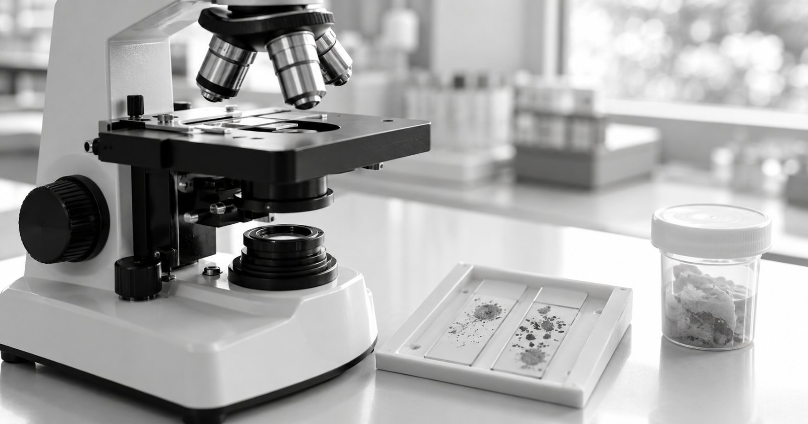
Cytology examines cells collected from a lump or lesion. A biopsy provides tissue for histopathology, which examines cells within their surrounding structure. These investigations answer related but different questions. Your veterinarian chooses the approach based on the examination, the location of the lump and what a result would change about the next step.
What can cytology tell you about a lump?
Cytology may help distinguish inflammation from a tumour and, in some cases, identify the type of cells involved. Samples can be obtained through aspiration or by making an impression from a suitable surface. The collection method is a clinical decision, not something to attempt at home.
The Merck Veterinary Manual's cytology guidance explains both the usefulness and the limits of examining individual cells. Some lesions shed too few representative cells for a clear interpretation. A report may therefore describe findings without giving a definitive diagnosis.
How is a biopsy different?
Histopathology allows a pathologist to assess tissue architecture as well as the cells. A biopsy may be needed when the clinical question depends on that structure, or when cytology does not explain the lesion adequately.
The word “biopsy” does not itself mean cancer. Tissue examination can also investigate other disease processes. Likewise, a lump's appearance alone cannot establish its cause. The Merck guide to diagnosing skin disease describes the importance of history, examination and selecting appropriate diagnostic methods.
What does an inconclusive cytology result mean?
An inconclusive result means the submitted material has not settled the question. It is not the same as a reassuring diagnosis, and it is not proof of malignancy. Ask your veterinarian whether the report recommends a different collection approach, tissue examination or another investigation.
A useful follow-up question is: “Does this result fit what you found on examination, and what information is still missing?” Keep the complete report, including comments, for that discussion.
What information should accompany a sample?
For a clinic submission, record:
- The animal's identifier, species, breed, age and sex.
- The precise sampling site, with separate identification if there is more than one lesion.
- How long the lump has been present and any observed changes.
- Relevant treatment, previous results and the reason for sampling.
- The question the veterinarian wants the laboratory to answer.
Cornell's clinical pathology submission guidance stresses patient identification, sampling site and clinical history. Labels and paperwork should describe the same animal and lesion.
Before arranging a submission to eLAB Diagnostics, the clinic should confirm the investigation and specimen requirements using information for veterinarians or call 1800 889 1900. This article does not confirm current test availability. Collection and transport instructions should come from the receiving laboratory.
Frequently asked questions
Can cytology and biopsy be used for the same lump?
Yes. They can contribute different information during an investigation. Ask what additional question the second test is intended to answer.
Does every lump need both tests?
No single approach suits every lesion. The veterinarian should explain the proposed investigation and how its result will guide the plan.
Can I tell whether a lump is harmless by looking at it?
Appearance alone does not establish a diagnosis. Arrange a veterinary assessment and describe any changes you have noticed.
What should I do after receiving a laboratory report?
Review the full report with the treating veterinarian. Ask whether the findings match the examination, whether any limitations remain and what follow-up is recommended.